DEPARTMENT OF ORAL PATHOLOGY WORKSHOP ON PRP & PRF - 29.07.2024
TAGORE DENTAL COLLEGE & HOSPITAL
ORAL AND MAXILLOFACIAL PATHOLOGY & MICROBIOLOGY
ABOUT US
Department of Oral and Maxillofacial Pathology is been conducted in a meticulous manner. We have been equipped with the state of art equipments. Our department has well-trained teaching staffs. We have an excellent auxiliary staff for the purpose of biopsy processing, special staining and basic haematology. We have a systematic approach in moulding the students right from I year and we play a role not only in teaching by different methods but, making them a good human being and a knowledgeable individual.
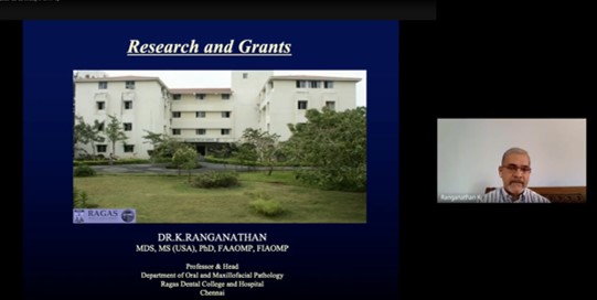
Students have secured 3 university gold medals for the subject Oral Pathology and Microbiology from The Tamil Nadu Dr.MGR Medical University for the academic year 2014, 2015 and 2019. Our department is involved in 2 ICMR projects with one being completed in the year 2014 and another being conducted in collaboration with Department of Pharmacology, Tagore Medical College and Hospital. Department holds a total of 53 publications in various International and National journals. Various research projects are being done by staffs and students in the field of Forensic Odontology, Oral Pathology and microbiology. We have conducted CDE programmes on various topics and in association with department of Prosthodontics and Conservative dentistry, we have been conducting Preclinical skills competition every academic year.
Oral Pathology deals with the nature of oral diseases, their causes, processes and effects. It relates the clinical manifestation of oral diseases to physiologic and anatomic changes associated with these diseases. Department includes lab, museum, A.V. room with L.C.D projectors, histopathology lab and microscope section. It has facilities to conduct various histopathological examinations and also a complete haemo
WORK DONE
Oral and Maxillofacial Pathology and Microbiology - Clinical Lab
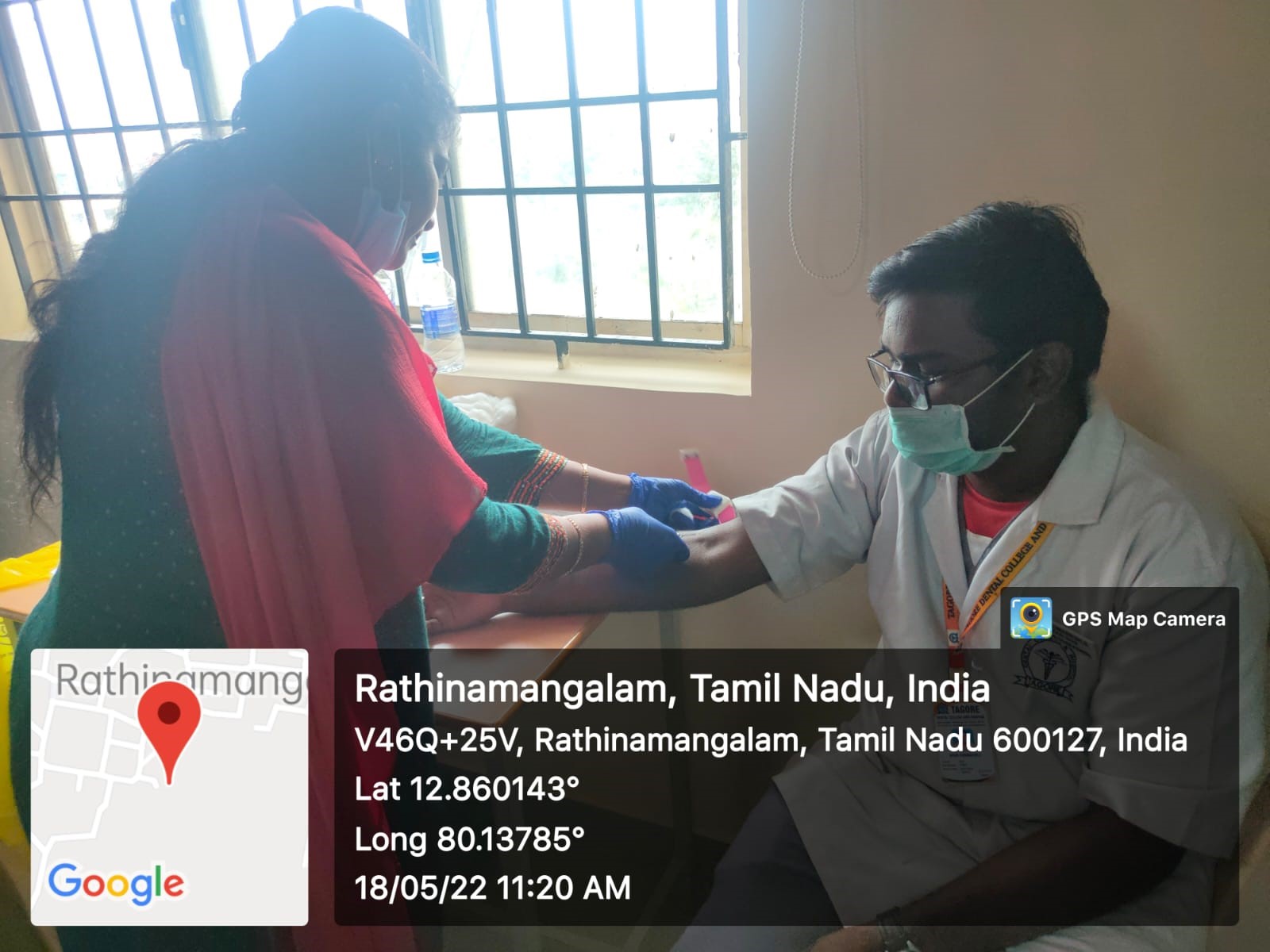
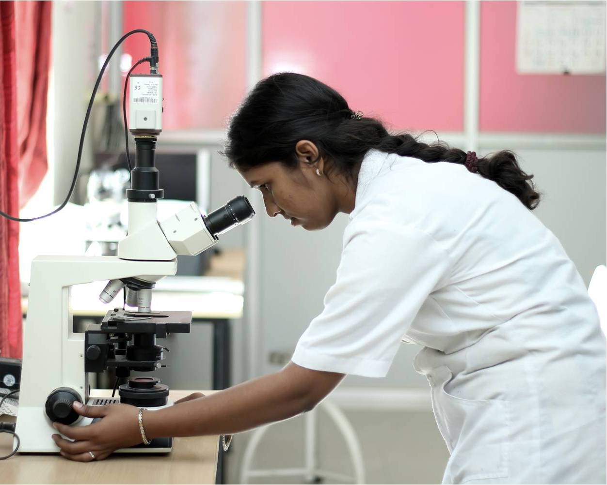
Our lab receives adequate number of Biopsy and cytological specimens for Histopathological examination. We have advance Research Microscope with conventional, polarising, dark field modes and a camera to photograph the slides, which can be mailed to the clinicians and can be stored for the purpose of future reference. Based on our report, treatment for the patient is decided. The lab is also equipped with micrometry devices for carrying out research which require measurements. Colorimeter device which is available in the department is employed in regular blood investigations and also for research purposes to estimate the serum, salivary levels of various components.
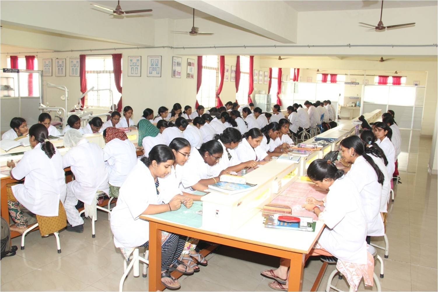
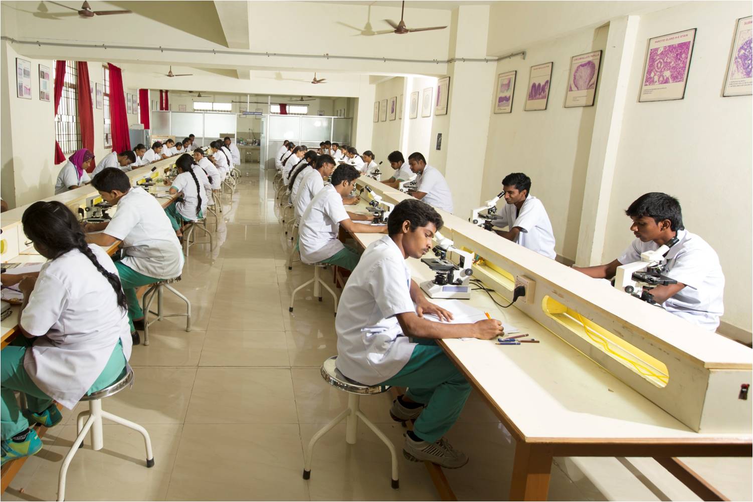
Oral and Maxillofacial Pathology and Microbiology - Students Lab
U.G. lab has a museum covering a wide range of specimens, A.V. room with L.C.D projectors, and a contemporary histopathology lab, a microscope section with illuminated monocular and binocular microscopes, and trinocular microscope with LCD projection.
TEACHING AND LEARNING
FOR INTERNS
The department of oral and maxillofacial pathology in the aim of producing good dentist who are sound in basics and pathological knowledge has formulated a curriculum which focuses on both clinical and practical exercises. The interns for each academic year attend a 15 days regular posting and as an elactive posting in the oral pathology department. During the period of posting, the interns areenlightened about basic protocol and disciplinary action to be followed in the department. The students are given instruction regarding attendance, quota and cases to be done. As a part of internship curriculum, the interns have to complete one long case, one short case, ground section ( Permanent longitudinal and cross sections, deciduous longitudingal sections), seminars and projects. The clinical cases screened by the interns are presented as a powerpoint seminar followed by a discussion between staffs and interns. The students have to completed a set of ground sections which they are allowed to interpret on their own in the microscope and followed by a discussion with the staff.
The interns are allotted a topic for seminar presentation based on BDS syllabus which is to be present in the seminar clubs. They also have to prepare models or charts based on topics and which is well preserved in department museum for further academic use in theory and practical classes. The interns are also given a overview on haematological exercises and also assist, perform basic haematological investigations like Hb estimation, Bleeding and clotting time.Interns have regularly posted to the department of oral medicine for screening of oral lesions, which they have record in the special cases registers which maintains the details of all lesions with regards to habits, frequency and various other parameters. After screening of the patients, they have to collect oral cytological smears which is transported to the department in a fixative and later stained with hematoxylin and eosin by the students themselves and interpreted for the diagnosis. Others specials stains like PAP, PAS staining procedures are also taught during their posting.
At the end of the posting, the interns are instructed to submit all the works in a folder as hard and soft copy along with ground section submitted and signature of the staff to be received in log sheet, record note and department registers. After final approval, the interns have to get HOD signature in the log sheet and record note and evaluation sheet with HOD signature to be submitted to college for course completion procedures.
FOR FIRST YEAR
Department of Oral and Maxillofacial Pathology which handles the oral histology for the first years in view of making the first year students strong in the basics of embryology, histology and tooth morphology handles various strategies to upgrade the knowledge of the students.
At the beginning of the academic year, initial orientation to the subject is given to guide between the transition period from school to professional courses. Basic topics to embryology and histology are covered in the theory and corresponding microscopic slides are shown to them in practical classes. At the end of each theory topics students to given tests as question and answer, diagrams, viva voce and other creative methodologies.With regards to practical aspect, the histology practical classes are conducted once in a week. Microscopic slides corresponding to theory topic that is in progress is focused in microscope to students which can be well correlated with theory knowledge that is perceived and for better understanding of the topics. Histologic pictures of slides seen in microscope with labelling are projected in the LCD television for students to follow up when they view the microscope. Hand digrams are foucsed in OHP projector for the entire class which to be drawn using H&E pencil. After staff approval in observation, they are transferred to the oral histology records.
Practical class also focuses on tooth morphology, where students are given through knowledge on types, chronology of dentition and tooth numbering system before carving classes. After acquiring basic knowledge in tooth morphology, the staff members give a detailed practical demonstration of individual tooth carving along with videos played in the department television which they can follow while carving and carving videos are made available in the college academic portal which the student can access with their unique college email ID and also in youtube.
Tooth morphology practicals also focuses on teaching individual natural tooth with morphology, study models for age estimation which is a part of examination curriculum and also further enhances their knowledge before they enter clinical postings.The students are divided into batches for the entire academic year with individual staff allotted to them and they have to show all the histology and tooth morphology works to their respective staffs, get signature after completing each exercise and finally at the end of academic year, the staff incharge and HOD signature to be received in the record and submitted along with tooth carving arranged in a neat labelled box for the final university examination.
FOR THIRD YEAR
Department of Oral and Maxillofacial Pathology, after imparting basic histological and embryological knowledge in first year, the third year curriculum is designed in view of making basics stronger and imparting histopathological knowledge. The theory classes are scheduled according to topics, of which few basic topics are covered in second year, so that major pathological topics are concentrated well in the third year to impart a sound pathological knowledge and which is the main aim of the department.
The practical classes are designed according to the corresponding theory classes which will help students to correlate the slides with the theory knowledge.Histologic pictures of slides foucsed in microscope with labelling are projected in the LCD television for students to follow up when they view the microscope. Hand diagrams are focused in the OHP projector for explaination and drawing the diagrams in the observation. After staff approval, to be transferred to the record.
Practical classes also focuses on demonstation of various study models depicting diseased state, which is to be recorded in the observation and being a part of examination spotters, the department aims in inculcating basic interpretation knowledge and applying it in clinical scenario when they step into dental practice for the welfare of the society.
The department conducts various practical exams to increase the familiarity with histopathological slides, viva voce, creative drawing and model works, frequent theory tests to constantly keep them in studying phase and making a stronger pathologist, apart from clearing the university exams.
OUR TEAM

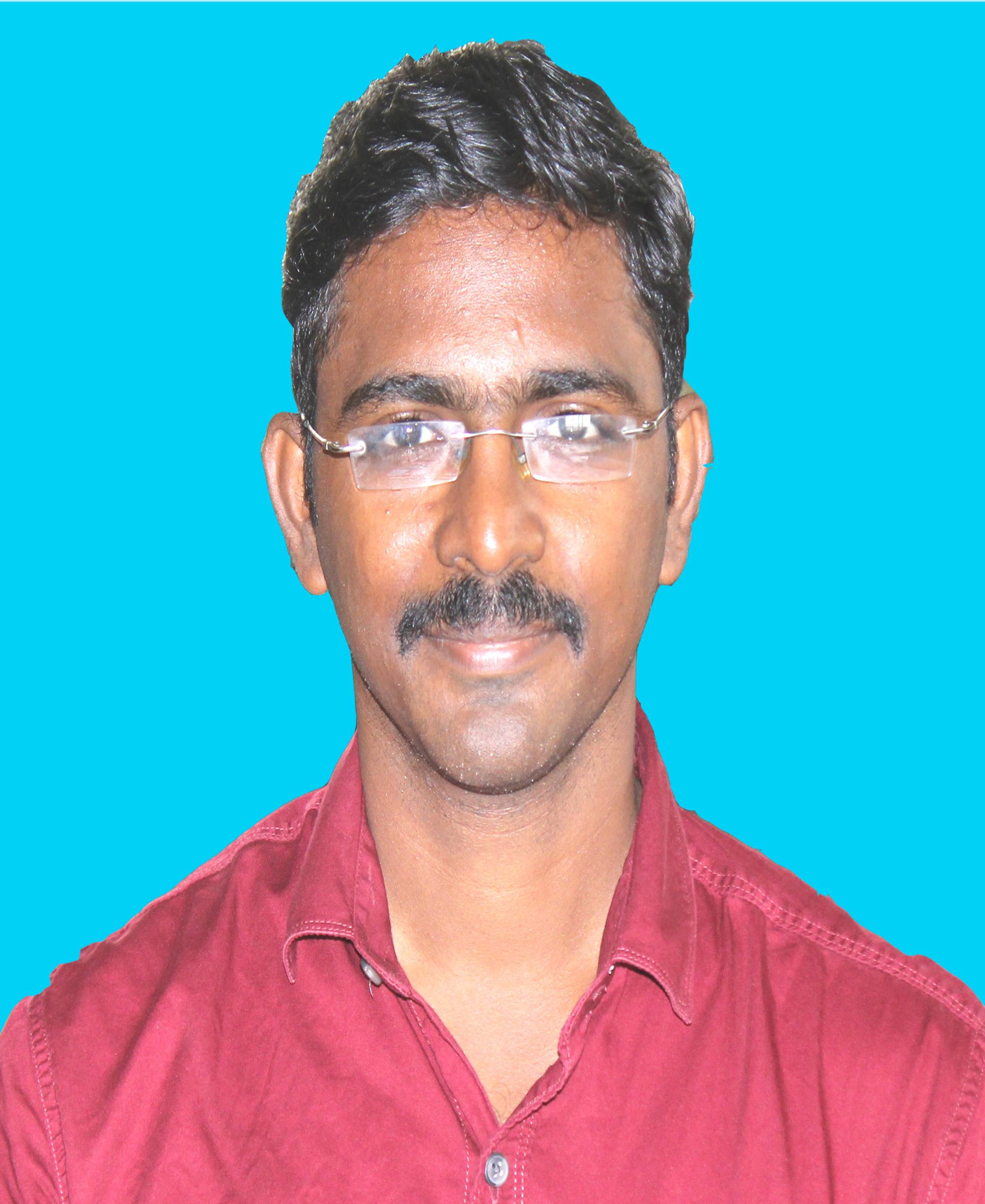
DR.P.SAI KRISHNA
PROFESSOR & HODHas a teaching experience of 22 years after MDS and 32 years of experience after BDS. He has 26 publications to his credit in various journals. He is in collaboration with Department of Pharmacology, Tagore Medical College and Hospital in an ongoing ICMR project. He was a former associate editor for JOMFP - An official journal for the Association of Oral and Maxillofacial Pathology. He was awarded with Fellowship from International College of Dentistry in December 2016. He is member of Instititutional Review Board. His topic of interest is in premalignancy and oral cancer
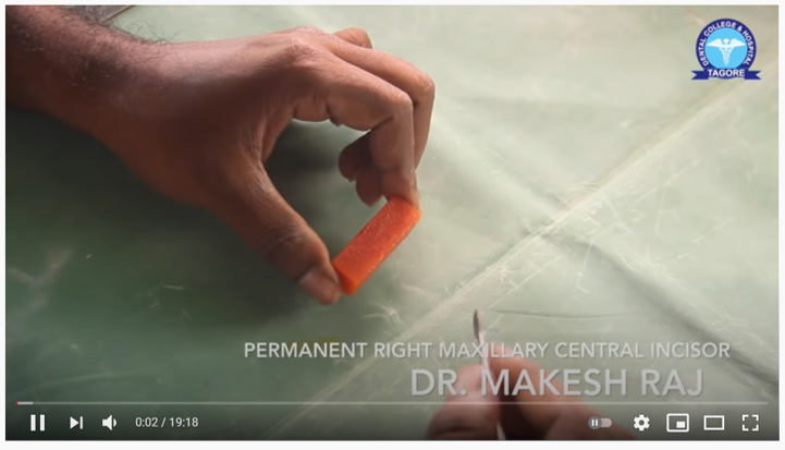
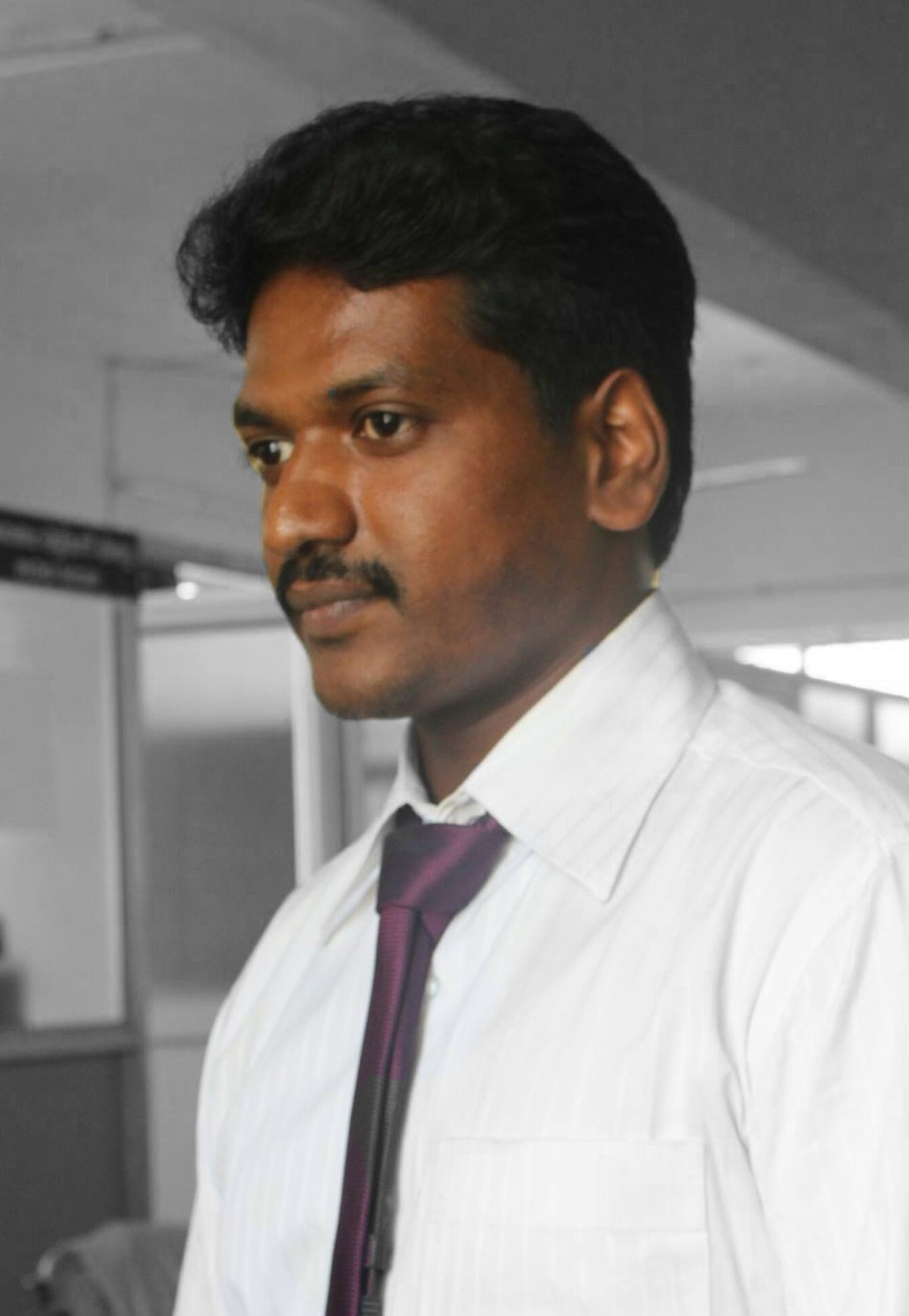
DR. L.S. MAKESH RAJ
PROFESSORHe has a teaching experience of 15 Years, 4 Months after MDS. To his credit he has 33 publications in various National and International journals and has contributed in 2 books for Undergraduate students. He has undergone PCR training programme in Sankara Nethralaya. His topic of interest is in Forensic Odontology and Oral Microbiology. He was awarded with distinguished faculty award for the year 2020 by Tagore Educational Trust.
DR. V. JAI SANTHOSH MANIKANDAN
READERHe has 9 years, 2 months of teaching experience. He received university gold medal for oral pathology in the year 2014 from The Tamil Nadu Dr.MGR Medical University. To his credit he has 16 publications in various National and International journals and authored 2 books. His topic of interest is bone and odontogenic lesions.
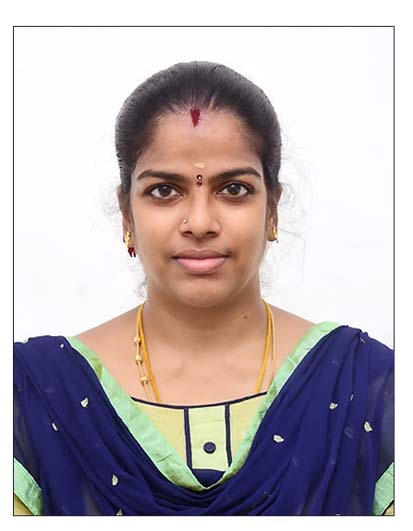
Dr.A.Hemalatha
Senior LecturerShe has 1 Year and 11 months of teaching experience. She has completed her MDS in Oral Pathology and Microbiology from SRM University in 2019. Has special interest in tooth morphology and forensic dentistry. She has 6 publications in national and international journals.
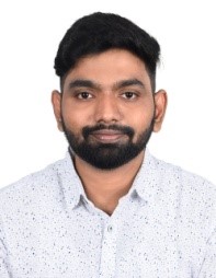
Dr. H. NANDA KUMAR
Senior LecturerHe has completed his MDS in Oral Pathology and Microbiology in the year 2023. He has 4 publications in indexed journals. His topic of interest is Oral squamous cell carcinoma and OPMDS.
DR. L. PRAVINA FERNANDO
LecturerShe has been associated with the department from 2006 with a total teaching experience of 17 Years 6 Months. She is involved in various departmental work and handling practical labs for first and third year. To her credit she has 3 publications.
SPECIAL EQUIPMENTS
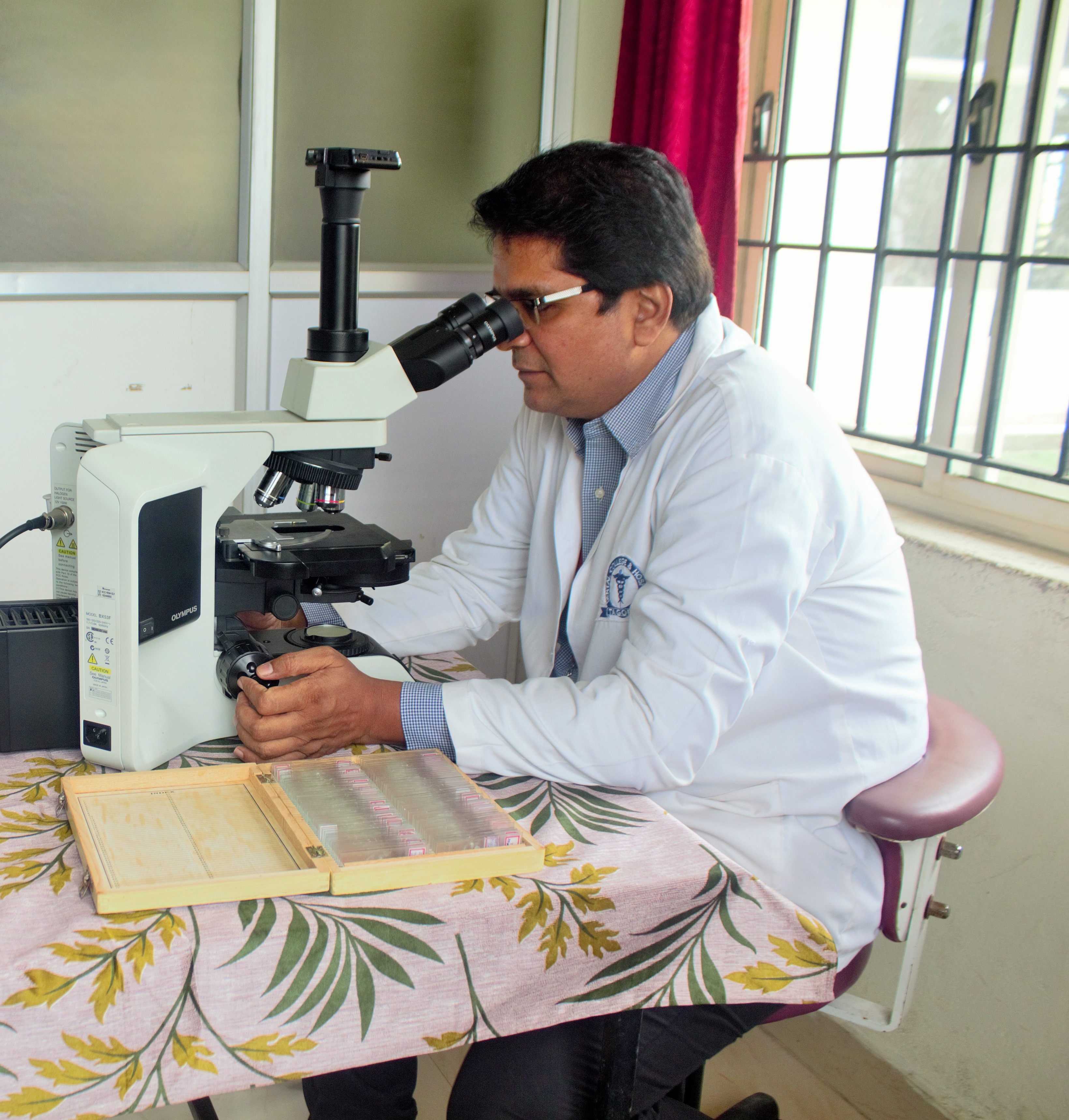
OLYMBUS BX 53 RESEARCH MICROSCOPEclick
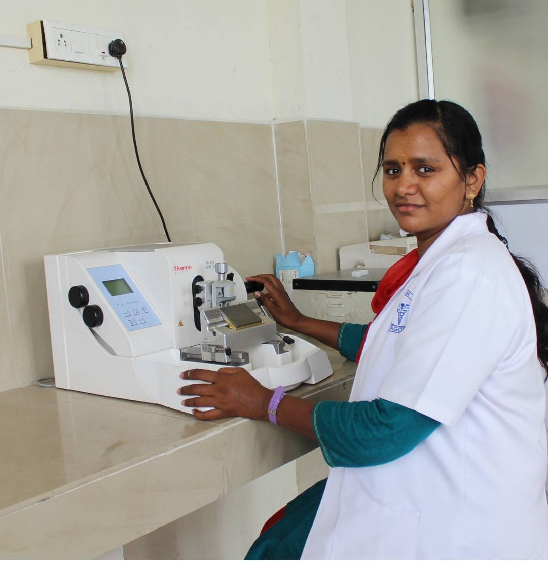
SEMI AUTOMATIC THERMO SCIENTIFIC HM 340E ROTARY MICROTOMEclick
EQUIPMENT LISTclick here
EVENTS & PROGRAMS